Home
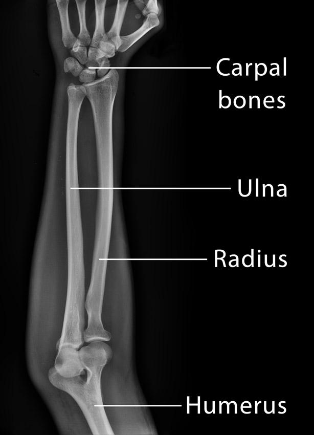
/ Radius Bone Labelled Diagram / Head, radial fossa, coronoid fossa, olacranon fossa, head... / The radius and ulna are the two bones of the forearm.
Radius Bone Labelled Diagram / Head, radial fossa, coronoid fossa, olacranon fossa, head... / The radius and ulna are the two bones of the forearm.
Radius Bone Labelled Diagram / Head, radial fossa, coronoid fossa, olacranon fossa, head... / The radius and ulna are the two bones of the forearm.. Learn the names by coloring. Radius anatomy pictures and information these pictures of this page are about:radius bone location. Labeled human forearm radius and ulna bone anatomy wall. Cheek bone (zygoma) upper jaw (maxilla). If you or someone you care for has sustained a fracture and are unable to.
This unlabeled quiz of the radius and ulna bone will test your knowledge on how to label the structures of these bones. Examples of long bones include the femur, tibia, fibula, long bone labeled : This tutorial covers basic features of the anatomy of the radius and ulna bones. The ulna articulates with the trochlea and the radius articulates with the capitulum. Bones of the left hand, view from below, labeled in latin.
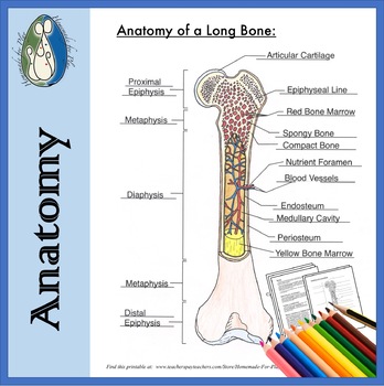
Bone Anatomy Diagrams for Coloring and Labeling, with ... from ecdn.teacherspayteachers.com Humerus labeled diagram stock illustration 181112825. It extends from the lateral side of the elbow to the thumb side of. Radius ulna bones ulnar notch anterior anatomy markings head forearm wrist hand limb upper pronation pivot supination during around. Drag and drop the pins to their correct place on the image. Each bone is a complex living organ that is made up of many cells, protein fibers, and minerals. The bones mentioned in each human skeleton chart are: The radius bone is the lateral bone of the forearm, and is homologous with the tibia of the lower limb. Learn everything about the anatomy of radius and ulna with our articles, video tutorials, labeled diagrams, and quizzes.
The tendon of the brachioradialis.
This tutorial covers basic features of the anatomy of the radius and ulna bones. Bones of the left hand, view from below, labeled in latin. Long bones are longer than they are wide and are the major bones of the limbs. Individually selectable every part, ideal for learning. The radius bone (os radius) supports the lateral (thumb) side of the forearm and the ulna bone (os ulna) supports the medial (little finger) side. In the medial surface, there. It lies laterally and parallel to ulna, the second of the forearm bones. All land vertebrates have this bone. I'm not sure of what you mean by bone diagram. Radius, in anatomy, the outer of the two bones of the forearm when viewed with the palm facing forward. The radius is considered the most commonly fractured bone in the human body, with distal radius fractures being the most common form of radial. Human body bone joint pains anatomy humerus with radius and ulna bones. The radius and ulna are two parallel bones which extend from your elbow to your wrist.
Learn vocabulary, terms and more with flashcards, games and other study tools. This long muscle travels nearly the entire length of the back. Drag and drop the pins to their correct place on the image. Learn everything about the anatomy of radius and ulna with our articles, video tutorials, labeled diagrams, and quizzes. The radius bone is this bone here and it lies laterally in the anatomical position.
Bone Anatomy Labeled Diagram Stock Vector (Royalty Free ... from image.shutterstock.com The ulna is usually slightly longer than the radius, but the radius is thicker. Its upper concave surface articulates with the. All land vertebrates have this bone. This long muscle travels nearly the entire length of the back. I'm not sure of what you mean by bone diagram. Radius anatomy pictures and information these pictures of this page are about:radius bone location. It is one of the two bones of the forearm, the other being the ulna. Lower jaw (mandible) collar bone.
Drag and drop the pins to their correct place on the image.
570 x 737 jpeg 46 кб. The radius and ulna together constitute the forearm. Drag and drop the pins to their correct place on the image. The radius bone is a long horizontal bone present in the forearm and is also called the radial bone. Illustration with skeleton of human hand ulna, radius and humerus bones. In its distal part, the radial shaft expands to form a rectangular end. The bones shown in the chest and hip region in the labeled human skeleton diagram are the ribs humerus is located in the upper arm. Labeled human forearm radius and ulna bone anatomy wall. 12 photos of the labelled diagram of radius bone. It extends from the lateral side of the elbow to the thumb side of. (ii) name the structure labelled a, which attaches muscle to bone. The ulna articulates with the trochlea and the radius articulates with the capitulum. The radius bone is the lateral bone of the forearm, and is homologous with the tibia of the lower limb.
Learn everything about the anatomy of radius and ulna with our articles, video tutorials, labeled diagrams, and quizzes. The three joints in the elbow. The bones mentioned in each human skeleton chart are: Almost every skeletal muscle works by pulling two or more bones either closer together or further apart. Drag and drop the pins to their correct place on the image.
Ulna: Definition, Location, Anatomy, Functions, Diagram from www.theskeletalsystem.net (vi) draw a labelled diagram of the cells as seen at high magnifi cation. A graphic shows the bones of the hand, carpals, metacarpals and phalanges. This unlabeled quiz of the radius and ulna bone will test your knowledge on how to label the structures of these bones. The radius is considered the most commonly fractured bone in the human body, with distal radius fractures being the most common form of radial. Radius bone is a photograph by asklepios medical atlas which was uploaded on august 3rd, 2016. This tutorial covers basic features of the anatomy of the radius and ulna bones. Bones of the left hand, view from below, labeled in latin. The radius and ulna are the two bones of the forearm.
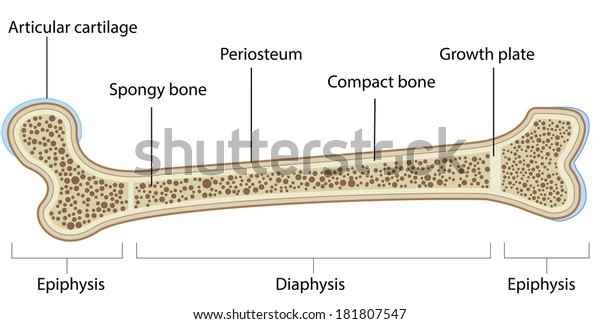
Long bones are longer than they are wide and are the major bones of the limbs.
(ii) name the structure labelled a, which attaches muscle to bone. Individually selectable every part, ideal for learning. 6 3 bone structure anatomy physiology the long bones are those that. The radius is a long bone in the forearm. It extends from the lateral side of the elbow to the thumb side of. The photograph may be purchased as wall art, home decor, apparel, phone cases, greeting cards, and more. Human body bone joint pains anatomy humerus with radius and ulna bones. This post discusses social security disability benefits and bone fractures. Projection of bone on the lateral surface of the distal radius bone. The bones mentioned in each human skeleton chart are: The radius bone is this bone here and it lies laterally in the anatomical position. Radius, in anatomy, the outer of the two bones of the forearm when viewed with the palm facing forward. The radius is considered the most commonly fractured bone in the human body, with distal radius fractures being the most common form of radial.
Start studying radius bone markings diagram labelled radius bone. Study guide for students and teachers.